Diagram Of Shoulder Bones / Arm Muscle And Bone Arm Bones And Muscles Diagram ... - The shoulder joint is the connection between the chest and the upper extremity.
Diagram Of Shoulder Bones / Arm Muscle And Bone Arm Bones And Muscles Diagram ... - The shoulder joint is the connection between the chest and the upper extremity.. Simple structure of the clavicle. Due to this, there is a hollow centre inside the backbone. Each vertebra has a hole in it. Following inferior dislocation of shoulder joint, the rounded contour of shoulder is lost and there is weakness of abduction of armbecause the. The largest bone in the human body is the thighbone or femur, and the smallest is the stapes in the middle ear, which are just 3 millimeters (mm) long.
Human shoulder diagram human shoulder anatomy stock photo anatomyinsider 129018944. Following inferior dislocation of shoulder joint, the rounded contour of shoulder is lost and there is weakness of abduction of armbecause the. The diagram below shows the anterior aspect of the scapula and its bony land marks. Bones have many shapes and sizes and are important to add structure to the body and protection to the vital structures. There also are bands of fibrous connective tissue—the ligaments and the tendons—in intimate relationship with the parts of the skeleton.
Guide to Shoulder Anatomy from embed.widencdn.net Download a free preview or high quality adobe illustrator ai, eps, pdf and high resolution jpeg versions. Shoulder joint is the most mobile joint of the human body. As a ball and socket synovial. Most shoulder fractures are treated successfully without surgery. It, essentially, floats off of the back of the chest, as it is connected to the body primarily by muscle. Human shoulder muscles and joints have a red signal. The largest bone in the human body is the thighbone or femur, and the smallest is the stapes in the middle ear, which are just 3 millimeters (mm) long. Shoulder bones and ligaments anatomy.
Related online courses on physioplus.
Ear wax normally comes out of your ear naturally so it's not a good idea to try and remove it yourself unless it is causing health problems (best to see your doctor first). Download a free preview or high quality adobe illustrator ai, eps, pdf and high resolution jpeg versions. There also are bands of fibrous connective tissue—the ligaments and the tendons—in intimate relationship with the parts of the skeleton. Shoulder problems including pain, are one of the more common reasons for physician visits for musculoskeletal symptoms. As a ball and socket synovial. It, essentially, floats off of the back of the chest, as it is connected to the body primarily by muscle. This framework consists of many individual bones and cartilages. The shoulder is the most movable joint in the body. The largest bone in the human body is the thighbone or femur, and the smallest is the stapes in the middle ear, which are just 3 millimeters (mm) long. Related online courses on physioplus. Consisting of the clavicle (collar bone) and scapula (shoulder blade), the pectoral girdle forms the attachment point between the arm and the chest. This lesson on the shoulder girdle, will complete the bones of the torso. Human shoulder muscles and joints have a red signal.
Posterior to the clavicle is the scapula, a flat, triangular bone located lateral to the thoracic spine in the dorsal region of the body. In humans they are triangular and lie on the upper back between the levels of the second and eighth ribs. The scapula, or shoulder blade, is an approximately triangular shaped bone. There are two kinds of cartilage in the joint. Shoulder joint of human body anatomy infographic diagram with all parts including bones ligaments muscles bursa cavity capsule cartilage membrane for medical science education and health care.
Shoulder Joint Ligaments - Medical Art Library from medicalartlibrary.com Shoulder joint is the most mobile joint of the human body. Scapula, either of two large bones of the shoulder girdle in vertebrates. In humans they are triangular and lie on the upper back between the levels of the second and eighth ribs. There also are bands of fibrous connective tissue—the ligaments and the tendons—in intimate relationship with the parts of the skeleton. Simple structure of the clavicle. This lesson on the shoulder girdle, will complete the bones of the torso. As a ball and socket synovial. Download 708 shoulder diagram stock illustrations, vectors & clipart for free or amazingly low rates!
Scapula, either of two large bones of the shoulder girdle in vertebrates.
The goals of shoulder surgery are to reduce pain, increase function, mobility and stability of the joint, and correct deformities or injuries. There also are bands of fibrous connective tissue—the ligaments and the tendons—in intimate relationship with the parts of the skeleton. These are the supraspinatus, infraspinatus, teres. This section will show images of the bones of the glenohumeral joint and their bony landmarks. The scapula, or shoulder blade, is an approximately triangular shaped bone. Bones » shoulder bones anatomy shoulder bone anatomy diagram human anatomy diagram categories: Shoulder bones and ligaments anatomy. The shoulder is not a single joint, but a complex arrangement of bones, ligaments, muscles, and tendons that is better called the shoulder girdle. This lesson on the shoulder girdle, will complete the bones of the torso. Posterior to the clavicle is the scapula, a flat, triangular bone located lateral to the thoracic spine in the dorsal region of the body. The shoulder is the most movable joint in the body. A normal, complete, human skeleton includes two shoulder blades, which the scapula is a flat bone so it is conveniently described by two diagrams, one to label the features of the posterior surface of the scapula and the. Due to this, there is a hollow centre inside the backbone.
Related online courses on physioplus. The shoulder joint (glenohumeral joint) is a ball and socket joint between the scapula and the humerus. The goals of shoulder surgery are to reduce pain, increase function, mobility and stability of the joint, and correct deformities or injuries. Shoulder joint is the most mobile joint of the human body. Human shoulder muscles and joints have a red signal.
Posterior view of the shoulder girdle bones - Netter ... from i.pinimg.com Shoulder joint is the most mobile joint of the human body. Human shoulder muscles and joints have a red signal. We discuss their function, the different types of bones in the human body, and the cells that are involved. The transverse humeral ligament is not shown on this diagram. Related online courses on physioplus. The shoulder joint is the connection between the chest and the upper extremity. The diagram below shows the anterior aspect of the scapula and its bony land marks. Scapula, either of two large bones of the shoulder girdle in vertebrates.
The rotator cuff muscles are four muscles that form a musculotendinous unit around the shoulder joint.
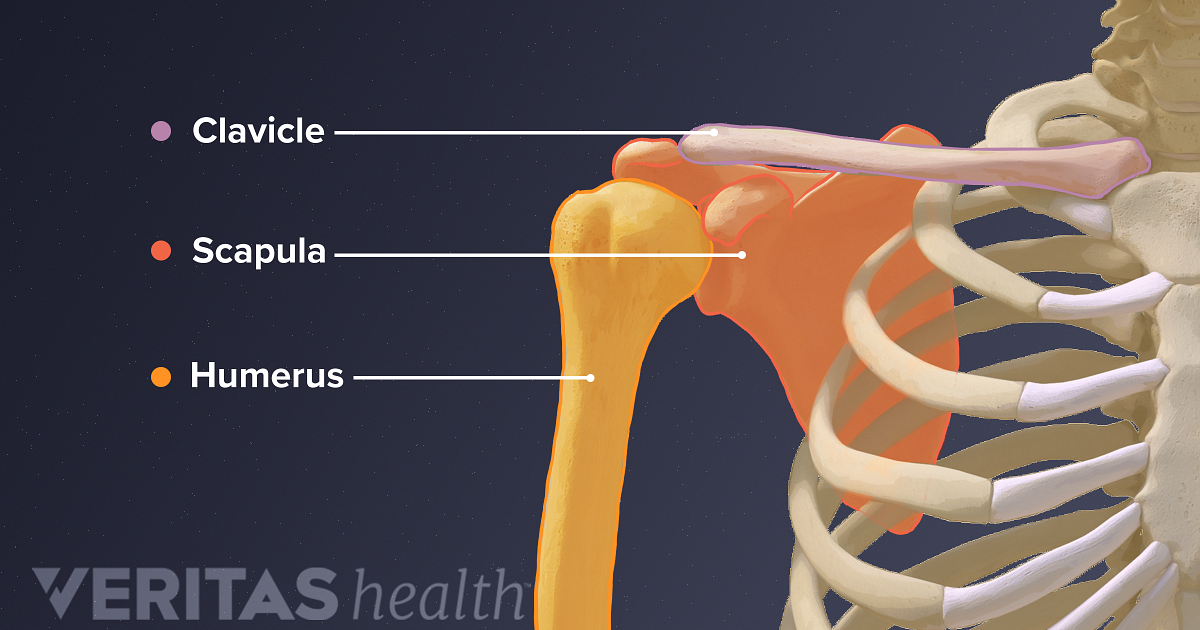
Related online courses on physioplus. (1) collar bone on the two sides of the next keep our shoulders apart. Shoulder bones and ligaments anatomy. The scapula is a large, flat triangular bone with three processes called the acromion, spine and coracoid process. In humans they are triangular and lie on the upper back between the levels of the second and eighth ribs. Consisting of the clavicle (collar bone) and scapula (shoulder blade), the pectoral girdle forms the attachment point between the arm and the chest. The transverse humeral ligament is not shown on this diagram. The bones of the shoulder consist of the humerus (the upper arm bone), the scapula (the shoulder blade), and the clavicle (the collar bone). Three bones come together at the shoulder joint. The shoulder bones, rib bones and hip bones ,are all joined to the backbone. The shoulder is a complex combination of bones and joints where many muscles act to provide the widest range of motion of any part of the body. This framework consists of many individual bones and cartilages. Human shoulder diagram human shoulder anatomy stock photo anatomyinsider 129018944.
There are two kinds of cartilage in the joint diagram of shoulder. Click now and learn everything about its anatomy and function at kenhub!